
Watery and Sticky Eyes

Overflow of tears shown by yellow dye

Narrowing of the tear duct opening (punctum), point shown by arrow (left) and by fluorescein (right)

Chronic infection of the canaliculus with punctal discharge

Watery eye with slow drainage of tears (note yellow dye) due to previous viral infection and canalicular blockage

Mucocoele of the lacrimal sac (note yellow dye failing to drain)

Lacrimal Examination (Dacrocystography, DCG), showing a patent right nasolacrimal (drainage shown by long arrow) and occlusion of the left duct (short arrow)

Lacrimal imaging (Dacroscintigraphy) showing blockage of tear outflow within the lacrimal ducts (arrows) despite normal clinical examination

Lacrimal drainage tube (arrow)

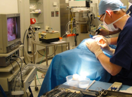
Placing a lacrimal drainage tube under GA
slide Next
slide